A New Technology Uncovers Very Old Secrets
The type of Computed Radiography (CR) that has been used to
take pictures of what has been preserved inside Leonardo was invented by
Eastman Kodak in Rochester, NY
in 1975. Since then, the technology has
been used mostly for industrial and medical purposes. Although to most people the images CR
produces look like the old familiar X rays we are used to seeing in doctor's
offices, there are some big differences.
The Leonardo science team used the industrial version of CR to look
inside the fossil. Because Leonardo had
turned to stone, the dinosaur was much denser than a living animal. Industrial CR is used for such purposes as
examining the concrete supports on bridges to see if they are cracked on the
inside. In order to see inside stone, it
is necessary to use much more powerful radiation that the kind of X ray that is
used in a doctor's office. To create the
radiation, three different types of radiation sources were used on Leonardo,
each one allowing the science team to get different quality images. The first was a water cooled X ray tube. This produced X rays nearly 5,000 times more
powerful than what you would get at your doctor's. Even with this high powered tube, each
picture took up to 40 minutes of exposure.
The second type was iridium 192, a type of radioactive material that was
much more powerful than the water cooled X ray.
Using this let the team look at thicker parts of the fossil and take
less time for each picture. The third
type was Cobalt 60, a highly radioactive source that was used to look at the
thickest parts of Leonardo. In fact, the
Cobalt 60 was so highly radioactive that even though it was used in a building
with lead-lined walls, the science team had to hide behind another building to
be certain they were not exposed to harmful radiation.
Computed Radiography doesn't use the same type of film that a regular X ray uses. It exposes a flexible phosphor plate. This looks like a thick piece of black plastic and in the case of Leonardo, it was placed directly on the fossil. The source of the radiation, either the X ray tube, the iridium or the cobalt, was placed opposite the film on the other side of the fossil. Then, the science team evacuated the building and the exposure began. In most cases the exposure time was between 20 minutes and one hour. When the exposure was finished, either the X ray tube was shut off or the iridium or cobalt was remotely placed back into a lead holder to protect the people in the area. At all times, a radiation safety officer had to be present and technicians were constantly walking the area with geiger counters measuring the radiation.
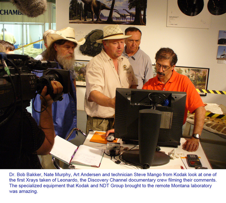
As soon as it was safe to approach Leonardo, one of the technicians would remove the imaging plate and place it in a machine that was a very specialized type of scanner. This machine used lasers to scan the surface of the plate and transfer the information from the plate to the computer. There, special software converted the scanned data into an image that looked just like a regular X ray. The difference between this image and a regular X ray is that there is much more information in the image and it can be manipulated by a technician operating the computer. It can be greatly magnified, brightened, darkened and filtered in ways that reveal a great deal. When it came to Leonardo, this amazing technology revealed much more than the science team hoped for. As the team gathered around the large monitor looking at the images taken at NASA's Johnson Space Center, Leonardo revealed that he had been severely bitten by a large predator shortly before he died. And two days later, the team looked at a dark mass in Leonardo's chest. Dave Trexler put it best as he said, "It's about the right size... It's in the right location... It could be his heart." (back to The Science)
Computed Radiography doesn't use the same type of film that a regular X ray uses. It exposes a flexible phosphor plate. This looks like a thick piece of black plastic and in the case of Leonardo, it was placed directly on the fossil. The source of the radiation, either the X ray tube, the iridium or the cobalt, was placed opposite the film on the other side of the fossil. Then, the science team evacuated the building and the exposure began. In most cases the exposure time was between 20 minutes and one hour. When the exposure was finished, either the X ray tube was shut off or the iridium or cobalt was remotely placed back into a lead holder to protect the people in the area. At all times, a radiation safety officer had to be present and technicians were constantly walking the area with geiger counters measuring the radiation.
As soon as it was safe to approach Leonardo, one of the technicians would remove the imaging plate and place it in a machine that was a very specialized type of scanner. This machine used lasers to scan the surface of the plate and transfer the information from the plate to the computer. There, special software converted the scanned data into an image that looked just like a regular X ray. The difference between this image and a regular X ray is that there is much more information in the image and it can be manipulated by a technician operating the computer. It can be greatly magnified, brightened, darkened and filtered in ways that reveal a great deal. When it came to Leonardo, this amazing technology revealed much more than the science team hoped for. As the team gathered around the large monitor looking at the images taken at NASA's Johnson Space Center, Leonardo revealed that he had been severely bitten by a large predator shortly before he died. And two days later, the team looked at a dark mass in Leonardo's chest. Dave Trexler put it best as he said, "It's about the right size... It's in the right location... It could be his heart." (back to The Science)